It was in 1987 that a lawsuit brought by 15-year-old Jenifer Graham of Victorville, California, U.S., resulted in several states offering an alternative to the dissection of frogs in science class. But for many middle school students, it is still a rite of passage and for good reason, according to David Evans, Executive Director of the National Science Teachers Association. Not only does the anatomy extrapolate to the human body, but it's hands-on: "There's something visceral and important about the real thing. What does this particular organ feel like? How stiff is it? Is it compressible?"
Showing posts with label dissection. Show all posts
Showing posts with label dissection. Show all posts
Saturday, February 28, 2015

 Posted with Blogsy
Thursday, November 20, 2014
Chopin's heart

The heart of Frederic Chopin has had a long journey (READ ABOUT IT HERE) since the death of the composer in 1849. At his request it was removed from his body, which was buried at Père Lachaise in Paris, and is now enshrined in a church in his native Poland. After it had been preserved in alcohol (possibly cognac) within a hermetically sealed crystal jar and encased in an urn made of mahogany and oak, it was smuggled into Warsaw by his sister. The organ was examined in 1945, when it was described as "incredibly large." A request by scientists in 2008 to test it and determine the musician's cause of death was denied by the Polish government. But in September, news broke that a group of 13 clergy and scholars clandestinely removed and examined the heart in the middle of a night in April 2014. The relic currently appears as an enlarged white lump submerged in an amber-colored fluid in a crystal jar. The team took hundreds of unreleased photographs, but were not allowed to take tissue samples. Poland's then culture minister Bogdan Zdrojewski was present and declares, "We in Poland often say that Chopin died longing for his homeland. Additional information which could possibly be gained about his death would not be enough of a reason to disturb Chopin's heart." Contradicting that remark is the official announcement by Poland that their native son's heart will be reinspected…but not for another 50 years.


Wednesday, September 18, 2013
Blob
The blobfish has gotten a lot of press since winning the vote as the ugliest endangered animal to become the official mascot of the Ugly Animal Preservation Society. But this is a blob of an entirely different sort. It is a human brain without any of the characteristic convolutions! People with this rare condition – known as agyria or lissencephaly – rarely survive childhood and are known to have muscle spasms, seizures, and a range of learning difficulties. This is the most profound example that neurologists have seen and would have remained unseen if not for Austin-based photographer Adam Voorhes:

"I walked into a storage closet filled with approximately one hundred human brains, none of them normal, taken from patients at the Texas State Mental Hospital. The brains sat in large jars of fluid, each labeled with a date of death or autopsy, a brief description in Latin, and a case number. These case numbers corresponded to micro film held by the State Hospital detailing medical histories. But somehow, regardless of how amazing and fascinating this collection was, it had been largely untouched, and unstudied for nearly three decades."The symptoms of this particular patient, however, are a mystery. The label on the jar states that he or she died in 1970, notes the anomaly, but includes a reference number corresponding to a microfilm which no longer exists…
THANKS TO WENDY MUIR FOR THE LINK!
Thursday, August 1, 2013
Pancras pinniped
"It came as something of a shock," said Phil Emery, the archaeologist with Ramboll UK, who led an excavation beneath the streets of London (IMAGE HERE) that revealed the 19th c. burial ground of St. Pancras Church (IMAGE ABOVE). It was not the quantity of bodies they found (1,500 of the 44,000 interred between 1822 and 1854 due to a wave epidemics) that was surprising. Nor was it the shared coffins, mass graves, and other evidence of hasty burial, which was understandable under the circumstances. It was the fact that in with a mix of human bones, they found the bones of a 13' (4 m) walrus! The reasons the animal was brought from the North Pacific to England, and where it had died, are a mystery. But signs that the walrus had been dissected - like the remains of a tortoise, rabbit, cat, dog, horse, and a couple of monkeys found during a recent excavation at the Royal London Hospital - point to curious medical students.

Friday, May 24, 2013
Disembodied tattoos

"You turn them over in your hands and you realise that they’re pieces of somebody else’s skin. Then you start to really look closely at the skin and think about all the associations you have with tactility and touch, and whether you can categorise them as objects at all. Because they have this power of subjectivity, still. A tattoo is a mark of somebody else’s will inscribed on their body, which in this case has outlived the individual, and it’s very, very strange to look at them and think about that."
These are the words of Gemma Angel, doctoral student at University College London. She has (for some) the enviable task of studying a selection of 300 tattoos acquired by the Wellcome Collection and now held in storage at the Science Museum archives in London. The specimens of inked human skin were purchased in Paris in 1929 from an osteologist and anatomist named La Vallete. They date from 1850 to 1920 and were likely obtained through the doctor's work at French military establishments and prisons, but the identities of their bearers remain unknown. In fact, not much is known about the collection at all and the tattoos have never been comprehensively studied. Angel's background as a tattooist gives her some insight as she explores the material properties of the skins as prepared specimens, the iconography of the tattoos, and the social and historical contexts of their preservation and collection.
Friday, April 27, 2012
Tim Curry as Alexander Monro!
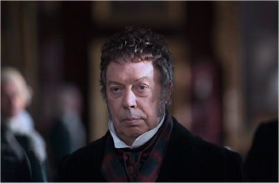
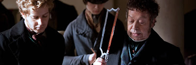

Monday, March 5, 2012
Dissection on display

My book has been published! Dissection on Display: Cadavers, Anatomists and Public Spectacle is a good read, if I don't say so myself. Here is the last paragraph of the Preface:
"The dissectors of earlier times and today have had the ability to mold bodies into anatomical marvels curated for future generations in medical museums, or to hold an audience spellbound by cutting cadavers apart. What these bodies symbolized for the anatomists and those whoIf this piques your interest - morbid, medical, historical, or otherwise - the book can be ordered (like my previous 5) directly from McFarland. It is also available from Amazon. Those of you who have followed along during my 3-year research and writing process* know my inspiration and the dedication it took to bring the project to fruition. I can now say I am pleased with, and proud of, the result!
saw their work—during their lifetimes or long afterward—had much to do with the age in which they lived. During the Renaissance, Europeans crowded into newly-built anatomy theaters to see for themselves how the hand of God manifested itself in the internal workings of the human body. The revolutionary anatomist Andreas Vesalius was teaching those who watched in such an engrossing way that knowledge of anatomy came firsthand, not at arm’s length through the published mistakes of predecessors. Asthe history of anatomy unfolded in the eighteenth and early nineteenth centuries, throngs of commoners clamored after an execution to ensure that the sentence to be hanged and publicly dissected was carried out to its ignominious end. The lesson to the audience, as depicted in William Hogarth’s famous illustration, was that crime does not pay. The moral took the form of the undraped corpse picked apart in front of friends and neighbors—and perhaps skinned to make manifest the metaphorical book of the body. For Victorians watching the much-anticipated unwrapping and examination of an Egyptian mummy, Thomas Pettigrew was dissecting another culture for his well-heeled audience, not merely pulling apart an ancient and exotic human corpse. But early in the twentieth century, public dissection went underground. The cadaver came to signify privileged knowledge as witnessing and participating in dissection became the prerogative of doctors and doctors-to-be as part of their professional development. To revisit how the doors of the anatomy theater were opened to the public, closed to those without credentials, and then thrown open again, let us take a seat as the anatomist takes the stage..."
*during which the blog byproducts included posts about career and artistic anatomists, an anatomy theater, the dissection of animals (an elephant and a squid), notorious bodysnatcher William Burke and what was done to his body, anatomical paintings by Rembrandt and modern artists, and dreams about dissection by Freud and a French scholar.
Friday, January 20, 2012
Burke & Hare


 On January 28th, for the 1st time in its 127-year history, the Anatomy Museum at the University of Edinburgh will open its doors to the public - and will continue to do so from 10am-4pm on the last Saturday of every month. The date is of particular importance, since it is the 183rd anniversary of William Burke’s execution. If you know about the history of anatomy, the name will need no further explanation, and is forever linked to his accomplice William Hare. The pair are infamous 19th c. bodysnatchers who committed at least 15 (and possibly as many as 30) murders for the money they could make supplying Scottish anatomists, including Dr. Robert Knox (1791-1862), immortalized in rhyme. While most of their fellow "resurrectionists" were exhuming newly-buried bodies from their graves to be used as cadavers, Burke and Hare found it easier to lure victims to their deaths (by smothering) and thereby a) ensure the freshness of their products and b) avoid the mess entailed in harvesting them from the ground. Though equally culpable for the appalling crimes, the 2 Williams suffered very different fates and are therefore represented by contrasting specimens in the museum:
William Burke
(1792- 1829)
After arrest, Burke was tried in Dec. 1828 for the murder of one of the victims, found guilty, and sentenced to death and public dissection. “Burke's body is to be dissected, and his Skeleton to be preserved, in order that posterity may keep in remembrance his atrocious crimes,” read the handbill that circulated at the time. He was hanged before a crowd of 20,000 on Jan. 28th, 1829, and his body was brought to Dr. Alexander Monro tertius (1773-1859), Dr. Knox’s rival. During his dissection, the anatomist made a death mask, the cast of which was recently found in storage at Inveraray jail in Argyll (1st image, held by Director of the Anatomy Department Dr. Gordon Findlater). After some 24,000 people filed past Burke's mutilated body, his skeleton (2nd image) was processed and his skin was tanned so that it could be crafted into souvenirs.
William Hare
(1792/1804 - ?)
Because there was insufficient evidence to secure a conviction against he and his partner, Hare was convinced to turn King's evidence and testify against Burke. His fate after his release is unclear. Some believe he left Edinburgh for Dumfries and was afterward seen in Carlisle. Others think he made his way to London, was blinded after being thrown into a lime pit, and spent the rest of his life on the street as a beggar. All that remains is speculation, an illustration, and a life mask (3rd image).
Those curious about the history of anatomy and dissection may wish to preorder my 6th book, Dissection on Display, which has now been announced for publication this spring/summer!
Monday, December 12, 2011
Shoe leather


When I was studying anthropodermic books in graduate school, I came across related examples of items other than book-bindings made from human skin. Among them was this pair of shoes (2nd image, color photo here) fashioned from the hide of "Big Nose" George Parrott. Thanks go to Sue for bringing the story back to mind so that I can relay it to you. It will not be my 1st post about shoes (see Burial shoes, Klompen, and Gumshoe), but it may be the strangest...
George Parrott a.k.a. George Manuse and George Warden was an American cattle rustler. In 1878, after he and his gang bungled a train robbery in Wyoming, they murdered 2 lawmen, resulting in a $20,000 bounty offered for their apprehension. The following year, still at large, they pulled off the daring daytime robbery of as much as $14,000 from wealthy Montana merchant Morris Cahn - despite the fact that Cahn was traveling with more than 2 dozen army troops. Still in Montana, Big Nose George was arrested in 1880 and returned to Wyoming to face charges of murder. He almost managed to escape from jail days before he was to hang, and an outraged lynch mob some 200-strong strung Parrott up from a telegraph pole. The doctors who took possession of the body after death used its components for a number of purposes:
- They studied the outlaw's brain for clues to his criminality.
- They sawed off his cranium and presented it to Lillian Heath, who later became Wyoming's 1st female doctor, and is said to have used it as an ashtray, a pen holder, and doorstop.
- A death mask was made of Parrott's face.
- The skin of his thighs and chest was removed and sent to a tannery in Denver, where it was made into a medical bag and a pair of shoes that one of the doctors wore to his inaugural ball when he was elected governor of Wyoming in 1892.
"I was incredibly lucky to visit the day that photographer Craig Pindell and historian/writer Larry K. Brown were photographing George for the archives of the Wyoming State Museum. After they were done, they put the shoes and skull back in the normally closed-up glass case. When they weren't looking, I managed to cop a feel of the shoes, which are 2-toned. They're skin color on the front and brown in back - kinda like saddle shoes. Only the soles are made of cow leather. And they are very worn, proving that the doctor wore his George shoes a lot. I rubbed the shoes. I figured they'd be flimsy. I was shocked that they were very sturdy."The shoes, partial skull, and death mask are on permanent display at the Carbon County Museum (photos here and here). The skull cap may be seen at the Union Pacific Museum. The medicine bag has never been found...
NOTE: If you are hungry for another instance of shoes made from human hide, have a look at this 1883 report about the alleged gruesome activities that took place at the Tewksbury State Almshouse in Massachusetts. Among other abuses, the lengthy report details the tanning of human hides and includes patterns for cutting the leather and a photograph of a sample that was entered into evidence.
Wednesday, September 28, 2011
PSA
If the video doesn't play, click here.
The state of Florida ranks 18th within the U.S. for the prevalence of cigarette smoking among adults. We have over 2,509,000 current cigarette smokers aged 18 and above. The statistics in this country range from 9.3% in Utah to 26.5% in West Virginia (you can check out your state here, or compare statistics for your country if you are outside the U.S.). In an effort to reduce the number of smokers by 100%, and to prevent nonusers from starting to use tobacco, Tobacco Free Florida has been running some very impactful public service announcements. One very moving spot, which doesn't appear on their YouTube channel, features a man explaining that his wife died of lung cancer at 40 and closes with his words, "I never thought of 20 as middle-aged." Two more recent spots (here and here) focus on the physical effects of the inhaled smoke and a third (above) - which has got to be one of the most outrageous PSAs of all time! - concentrates on just one harmful ingredient...
Monday, January 10, 2011
Fortune's bones

I heard about this story in a book I just reviewed which mentioned a collection of poems for kids by Connecticut’s Poet Laureate Marilyn Nelson (excerpt above). Fortune (c. 1740-1798) was the head of a family of 5 slaves - including his wife Dinah, their son Jacob (b. 1786), and their daughters Mira (b. 1789) and Roxa (b. 1791) - owned by Waterbury physician Dr. Preserved Porter. Fortune and family lived in their own house on the Porters' 75-acre farm, which produced rye, Indian corn, onions, potatoes, apples, beef, hogs, cider, hay, oats, and buckwheat. Fortune most likely managed the farm so that Dr. Porter could attend to his medical practice, and Dinah would have cleaned and cooked. A 19th c. source indicates that Fortune slipped on a rock, fell into the Naugatuck River, and drowned. After the accident, Dr. Porter prepared Fortune’s skeleton to use as a teaching aid in the anatomy classes he taught. Fortune's wife and his skeleton remained the property of the Porter family. When Dr. Porter died in 1804, Dinah was inherited by his widow and the skeleton was inherited by his son Jesse Porter, also a doctor. It passed on to generations of Porter physicians until donated to the Mattatuck Museum in 1933. Fortune’s
"We are brief incarnations,
we are clouds in clothes.
We are water respirators,
we are how earth knows.
I bore light passed on from an original flame;
while it was in my hands it was called by my name.
But I am not my body,
I am not my body."
 articulated bones were first placed on exhibit in the 1940s, contextualized within the history of medicine and slavery in Waterbury by the 1960s, and removed from display as the exhibit of human remains became politically sensitive in the 1970s.
In the 1990s, the Mattatuck Museum began to search for reliable evidence about Fortune's life and death. "Fortune lived in the doctor's household and was part of the family and was loved. But one day Fortune is at the Naugatuck River and he slips in and drowns. The story has it that Dr. Preserved Porter jumps in to save him, but can't. And so he flayed his body on the spot and took him home. Would you do that to the servant you love as part of the family?" asks anthropologist Dr. Warren Perry (2nd image). He and other anthropologists and archaeologists examined the skeleton, which belonged to a man of about 60 years of age in relatively good health. The rugged bone structure and muscle attachments suggested that Fortune was a powerful man accustomed to the heavy work of a farmer. Healed injuries indicated that he had broken his lower back and damaged the bones in his hands and feet, which would not have been unusual for an agricultural worker. A snapped vertebra points  to a cause of death by a sudden jerking back of the head, but there was no additional damage to suggest a hanging or a fall. In addition to the visual examination, the bones were measured and x-rayed (which offered probable height, age and illnesses), and DNA analysis (which may identify his descendants) and chemical analysis (which may determine where he grew up) were performed. A facial reconstruction prepared by forensic artist Frank Bender and a full-figure portrait (3rd image, cropped) painted by medical illustrator William Westwood have become the centerpiece of the museum’s exhibit about Fortune and the African-American community in Connecticut in the 18th c. Despite the value of the 200-year-old remains remaining above-ground and uncontaminated so that they will be available to researchers as future methods of analysis are developed, the question of whether to bury Fortune's skeleton has inevitably followed.
Sunday, January 9, 2011
Compare and contrast


These two weird news stories surfaced a week apart last month:
Medical students at Rende Medical College in Taiwan undergo a lesson intended to give them an insight into death. They each write a will, dress in a funeral shroud, and climb into a coffin which is lowered beneath the floorboards (2nd image). Professor Qiu Daneng explained, "Although it's just 10 minutes, the effect is equal to real death." Student Xiao Lin concurred and said that it taught her that every second counts in life.
Medical students at the Karolinska Institute in Sweden recently underwent a life lesson. When they began their first clinical autopsy, described as an intense and trying experience, they learned that the cadaver they would be working on was the body of their former instructor. Although the name had been told to the class in advance, it did not register with the students until they read it on the corpse's toe tag. The tension rose and the group whispered among themselves, but nobody spoke up when the autopsy technician got to work. The incident did weigh on them afterward. "The first autopsy is very emotional and we autopsied someone we knew," said a student. "It was extremely unfortunate. This is the first time I have encountered something like this," said Birgitta Sundelin, the professor of the course. "It is really terrible, but it is part of education sometimes. Unfortunately, they must deal with it," said Tina Dalianis, the head of the department. The president of Sweden's Medical Students Association, however, was outraged by the school's response, a reaction echoed by a medical student at another university: "Very unfortunate. Students should not have to feel uneasy during their education. It is important that an autopsy truly be the educational opportunity that it should be. The question is how much these students learned from the situation."
A little too close for comfort?
Wednesday, September 1, 2010
Dissecting an elephant


Here's something. I came across this passage last night while looking up details about British surgeon and anatomist Sir Astley Cooper (1768-1841). I quote from an 1843 biography by his nephew:
"In the course of the year 1801, an Elephant, which had been one of the principal features of the exhibit, died. Immediate notice of the circumstance was, as usual, sent to Mr. Cooper [2nd image], and, notwithstanding the unwieldy bulk and enormous weight of the animal, he determined to have it brought to his house in St. Mary Axe [4th image, a building in that London neighborhood], where he was still living, and to dissect it. He accordingly hired a cart, in which, after a considerable degree of exertion, the Elephant was deposited, being afterwards covered with a large cloth, in order that it might attract as little notice as possible on its way. In this manner it arrived at St. Mary Axe, and the cart having been driven into the court-yard before Mr. Cooper's house, the outer iron gates were closed, and they set about attempting to get it into an outhouse, devoted to purposes of dissection. All their efforts, however, to effect this proved unavailing, and after a vast deal of trouble, they found themselves obliged to leave it lying exposed in front of the building. During their attempts to remove the carcase into the dissecting-room, a large mass of persons collected outside the gates, and continued to watch their proceedings through the iron railing. The obstruction this crowd caused in the thoroughfare of St. Mary Axe was so great, that it was found necessary to cover up the apertures between the railings by throwing a carpet over them, and to move the body of the Elephant as far as possible out of the view of those who were passing in the street. They were enabled to do this the more effectively, inasmuch as the stabling projected before the dissecting-room, and the body being placed in the recess, was, by these means, to a great degree concealed from public view. Mr. Cooper was thus compelled to dissect this animal in the open air, and finding himself, from its enormous size, unable to perform the task alone, he invited several students from the Hospital [Guy's Hospital, 3rd image] to assist him. Here for some time they worked together, being barricaded from the observation of the public; nor did they cease their operations until they had examined every structure to the very bones. These latter were carefully prepared, and being articulated under Mr. Cooper's directions, formed the skeleton which, even up to the present day, may be seen in the Museum at St. Thomas's Hospital, - an existing memorial of my uncle's scientific enthusiasm."The dissection was quite a feat, as you can see - if you care to - in this National Geographic video. Note the presence of an audience in both centuries.
Thursday, May 27, 2010
The Dissecting Room, part II


We are not done looking at yesterday's image of a woman in a dissecting room. Michael Sappol reports that it did not originate with the 1898 book The Doctor's Window. Titled "Anatomy of the Heart" (or "She has Heart! or The Autopsy") it was an 1898 painting (1st image) by Spanish artist Enrique Simonet Lombardo (1866-1927) that was widely reproduced after Simonet (self-portrait, 2nd image) received several awards for it at international exhibitions. Although beautifully done, the black and white reproductions - including this 1906 photogravure - do not have the subtlety of the color image and its monochromatic hues broken up by the red heart held by the doctor and the blue bottle of formaldehyde in the window. The painting is on display at the Museum of Fine Arts in Malaga and is described as one of the most valued in its collections: "The anatomical study of the dead girl is flawless..." This article explains that the female subject is a drowning victim and the painter captures the moment at which the anatomist stares at the heart wondering about life's fundamentals.
Wednesday, May 26, 2010
The Dissecting Room
 The same friend who found the mutant dandelions gave me a framed copy of this lithograph before I left town last year. It hangs in my bathroom in a themed grouping that includes a page from an anatomy text, a photograph of Mr. Johnson demonstrating facial restoration technique to his students, and my collection of antique dissection photos. Curious as to the origins of the image, I tried to look it up online, but there wasn't much about it. Then I came across it while reading Michael Sappol's A Traffic of Dead Bodies as an example of 19th c. medical illustration that bordered on the pornographic. He sources it to a volume of poetry on medical themes that was edited by Ina Russelle Warren and published in 1898. The litho appears on p. 248 of the book, called The Doctor's Window, which also contains the poem "In a Dissecting Room" by Dr. William Burt Harlow:
Sightless eyes half closed beneath
Long, black lashes curling yet;
Wavy locks the pale face wreathe
With the salty drops still wet.
Lying there so silently
Womanhood reproachful seems;
'Tis a face that we may see
Reappear in troubled dreams.
Lifeless, wasted arm and hand
Stripped of skin by scalpel keen;
Shining tendons, band on band
Ligaments and muscles seen.
Wondrously the fingers move,
Answering to the testing touch
Of each muscle far above,
Whilst the learner marvels much.
Searcher, wouldst that thou could find
What mysterious power once moved
That dead form! How vain and blind
This long quest of ours has proved!
Now the forceps and the knife
Merciless attack the face
Eagerly with death at strife
Winning by a swifter pace.
Inch by inch the clinging skin
With reluctance parting shows
Unknown wonders far within,
Sources whence expression flows.
Tiny threadlike muscles here
Teach the lips to move in smiles;
Draw the eyelids tense with fear.
Close them when soft sleep beguiles.
These have knit the brows to frown;
Those have taught the mouth to kiss;
Care and pain have oft weighed down
Wrinkling forehead's calm with this.
These once spread the nostrils wide
When in anger breath came fast;
Or when blew from ocean's tide
Airs of health caught ere they passed.
Magic house, where sometime dwelt
Spirit, soul, howe'er 'tis known!
Ah, what thrills thy walls have felt!
Whither has thy tenant flown?
If this ruined home appear
Wonderful beyond compare,
What was then the dweller here
That could vanish into air?
Subscribe to:
Posts (Atom)


